Medical Imaging
Duration
4 years
Entry
Biomed & Graduate
Scope of practice
NZ and AUS
Cohort size
21
Medical Imaging
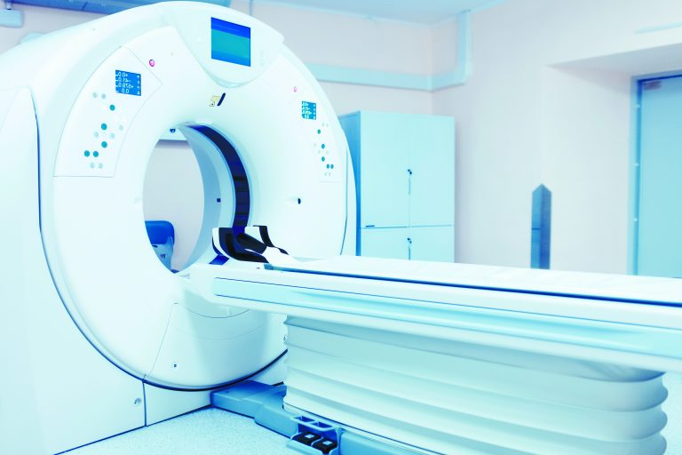
The Bachelor of Medical Imaging will prepare you to work as a medical imaging technologist. You will be imaging diseases and injuries using specialised equipment. Medical imaging technologists play an essential role since imaging is often needed to diagnose diseases and plan subsequent treatments.
As a medical imaging technologist, you will be dealing with a wide range of patients. You must have good communication skills since you will give patients instructions. Medical imaging specialities such as MRI, ultrasound and nuclear medicine require a postgraduate diploma.
Entry Statistics
Entry into medical imaging is competitive, and you will be selected based on your GPA (50% weighting) and interview (50% weighting). Nailing the interview is just as important as getting good grades! Below are some entry statistics to help you better understand the GPA needed for medical imaging. There are 23* seats for medical imaging, with first-year applicants taking up the majority of seats.
Want to maximise your chances? Check out our MMI Guide and receive a FREE Consultation today!
Core papers: BIOSCI 107, CHEM 110, POPLHLTH 111, BIOSCI 101, BIOSCI 106, PHYSICS 160, MEDSCI 142.
To be eligible for an interview, you must have an overall GPA of at least 5.0. Meeting this GPA will not guarantee you an interview offer. The interview cut-off GPA, calculated with core papers, is higher than this. This means that you need to do extra well on your core papers and maintain your grades in semesters one and two. The interview-cutoff GPA has been increasing over the past three years. In 2021, the interview-cutoff GPA was 7.0.
Similarly, to be eligible for an interview, you must have an overall GPA of at least 5.0. The GPA of the last two years of study is used for entry in the graduate pathway. The interview-cutoff GPA for 2021 is 7.3 and has increased over the past three years. This pathway can benefit those who did not do well in their first year of study.
In both categories, since the semester two grades are not available when interview offers are released, the highest possible grade is assigned for semester two.
This is a highly simplified guide. We strongly encourage you to visit the UoA medical imaging entry guide and student centre for more information. We try to make our information as accurate as possible. Still, entry criteria are subject to change, and our information may be outdated.
For entry tips, please visit our undergraduate and graduate guide.
Number of applicants
The number of applicants for medical imaging are dominated by undergraduate applicants.
No Data Found
Median successful core GPA
Although the median successful GPA is high, some students enter with a GPA much lower than this.
No Data Found
Programme overview & what students say...
Anatomy & Physiology
Anatomy and physiology relating to radiology
Patient care and saftey
How to perform imaging procedures safely
Hands on experience
Practical labs and placements
Specialist equipment
Learning how to use specialised imaging equipment
Part II
As a part II BMIH student, I did a mix of academic and clinical work throughout the year. In semester one, I did three MEDSCI papers which focused on Anatomy, Physiology, and Pathology and one MEDIMAGE paper which introduced us to the basic concepts of Medical Imaging. This included the clinical and professional aspects, as well as the ‘physics’ radiology content. In the second semester, I took four MEDIMAGE papers that further supplemented and added new information to what I had learned in semester one. Aside from the MEDIMAGE lectures, we had to attend clinical labs at Greenlane Clinical Centre and anatomy labs at the Human Anatomy Lab in the university.
The year was a mix of online and in-person sessions. It was challenging, but I very much enjoyed the experience. I was able to learn new things and gain new experiences. The clinical integration to the course was definitely the highlight of my year. I was able to practise the concepts I’ve learned from lectures and apply them to real life. It was a good balance of theoretical and hands-on experiences. Additionally, we had such a small cohort compared to Biomed (2020), which allowed our class to collaborate easily and establish a supportive environment. Finally, being able to meet new people from the upper year levels and clinicals added a bonus to my experience. Overall, it was a positive experience despite such an unstable year with the pandemic going on.
I would say that the workload and content are definitely a step-up from Biomed. The stage two MEDSCI papers that I took expounded on the content we tackled from MEDSCI 142 in first year Biomed. Additionally, that was the first year I took MEDIMAGE papers which specialised in radiology concepts. The lab aspect was interesting. The MEDSCI labs were more hectic than the first year since we had to write a full proper lab report rather than just worksheets. The MEDIMAGE and CLINIMAGE labs were very practical and also hectic since you have to process and understand a fair amount of content during and after each session. However, I would say that it is doable as long as you have good time management and a sound system of prioritisation.
As mentioned earlier, the content can be overwhelming, but it is manageable with a good and consistent approach. Some techniques that got me through the year were consistent revision of content, such as reiteration and active recall. However, the workload keeps getting heavier as you progress through the semester. Resorting to practice questions and past exam papers were very helpful. Long study hours does not guarantee excellent results. But instead, an effective study method is the key. I also found that building rapport with your classmates (as it is such a small cohort) is important. Last year, a few of my friends did mini-study sessions in between classes and we’d quiz each other. Lastly, I would highly recommend having a good work-life balance. Yes, hard work is important but taking breaks is more important. It allowed me to clear my head and prepare it to process more information. I went for long jogs in between study sessions or even just a simple walk to the park. Taking care of your well-being should be your priority because a well-taken care mind and body is a working mind and body.
Part III
Part III is where you solidify your knowledge in general radiography and begin to branch out from general x-ray. Your general radiography development will include learning about contrast media, adaptive trauma techniques, and x-ray specialisations (including fluoroscopy and paediatrics). You will be required to complete 12 weeks of placements, which will rotate between general radiology, emergency, theatre, interventional radiology, and more. In addition, the course will introduce you to other imaging modalities such as computed tomography (CT), magnetic resonance imaging (MRI), ultrasound (US), nuclear medicine (NM), and mammography. This journey will successfully start learning how to identify normal and abnormal anatomy/ pathology of 3D multi-slice CT scans, which will then carry you into CT in part IV.
The best part for me in my 3rd year was branching out to the other specialisations of imaging. The medical imaging degree only allows you to practice general radiography (x-rays) after you graduate; however, I was especially interested in what more you could do. Being introduced to the medical imaging specialties study taught me the basics about the other modalities I could take in the future. Learning about what they imaged and how they imaged expanded my interest and even solidified what my future might look.
Make the most of your social and university life in the first two years in medical imaging because your final year with around 30 weeks of placement will be like full-time work.
Part IV
Part IV is where you really get to consolidate all the theoretical and practical knowledge that you have gained over the past three years! You have fewer lectures with all academic learning done remotely, where you will explore the concepts of computed tomography (CT) and musculoskeletal trauma in more depth. You will then be able to use this knowledge in clinical practice during your 24 weeks of placements in general radiography and 6 weeks of placements in CT. Alongside this, you will gain insight into potential modalities that you may decide to specialise in later on, such as; interventional radiography (IR), magnetic resonance imaging (MRI), ultrasound (US), nuclear medicine (NM), and mammography. In addition to this, you will also be undertaking a research project in an area that interests you. By the end of the year, you will be ready to work as a qualified medical imaging technologist!
Placements can be quite stressful, especially as you are trying to develop your skills further whilst getting through your practicals and balancing other commitments. However, the clinical tutors are wonderful, they offer a massive level of support, and they are always happy to help!
One of the biggest challenges offourth year is balancing placements with practicals, your research project, other uni work and part-time jobs. Time management is crucial and having the support of your medical imaging friends is super helpful as they know exactly what you are going through!
What does the job involve?
Hospital settings
Imaging patients
Patient communication
Report writing
Medical imaging technologists are imaging experts who image patients for injury and disease. They prepare patients for scans such as CT and X-rays. Medical imaging technologists need to be skilled to produce the best images to assist with diagnosis and treatment. Medical imaging technologists also perform safety checks on imaging equipment and play a big role in patient safety since radioactive materials are used.
Medical imaging specialities such as MRI, ultrasound and nuclear medicine require a postgraduate diploma. Speciality programmes can be competitive but rewarding for those who pursue them.
For more information, CareersNZ has a good guide of what medical imaging technologists do.

Wonder what it is like being a medical imaging technologist? We asked one and here’s what they say:
What are some thing you enjoy about being a MIT?
“Working together with colleagues and other multidisciplinary staff. Depending on the environment, e.g. outpatients/in-patients/public/private, medical imaging can be a large team effort, especially when the patient is unwell and you need to acquire the images as efficiently as you can. (p.s. x-rays can be hard for patients as they need to hold still in a position which may be quite sore for them). You may work with radiology nurses, transit nurses, ward nurses, healthcare assistants, orderlies, Radiologists, ED doctors, other doctors as well as medical imaging professionals in other modalities.”
“I enjoy being able to use my skills to help patients in need and provide diagnostic imaging to help answer a clinical question. It also involves being creative and adapting your technique to patients presenting in any way, which can be challenging but very rewarding.”
What are some other opportunities with this degree?
“Medical imaging is such a large field with many opportunities available both in general practice and in the different modalities. As a new graduate, I find it very stimulating and marvel at all the different roles we play in the healthcare system.”
Medical imaging is a key component of research as a lot of research at the university is conducted using different modalities of imaging. You can work in different research organisations such as CAMRI and Auckland Bioengineering Institute at UoA. Part 4 Medical Imaging students are required to undertake a research project as part of the Honours component of the degree, so all the students will get some exposure in a research environment and dissertation/academic writing. Students are required to take ownership of their own projects with the individual supervisors and organise time for supervisory meetings, data collection, data analysis/presentation in addition to being on placement full time. The range of topics differs greatly (with some past topics involving the brain, spinopelvic alignment, the breast, and joint studies) with the projects using a variety of different imaging modalities (x-ray, mammograms, CT, MRI, USS, Nuc med).
The general scope of x-ray involves outpatient, in-patient, emergency imaging, fluoroscopic imaging, intensive care/post-op imaging and theatre imaging. There is also departmental imaging where the patient comes to you (the x-ray room) or mobile imaging where you need to go to the patient (usually the more unwell or post-operative patients). You can also take up a tutoring or management role (e.g. clinical specialist) within the department, which brings about further responsibilities and opportunities.
Mammography, Interventional radiology/angiography, Cardiac investigation, and Computed Tomography (CT) are further specialisations of medical imaging that still rely on the same radiation principles but require extra training and can include additional studies. PACS (picture archiving and communication system) is also an integral part of medical imaging and covers the storage and retrieval of all the images as well as tech support in this highly technical field.
Other specialisations in different modalities include nuclear medicine, ultrasound, and Magnetic resonance imaging (MRI) which require a post-graduate diploma (minimum 2 years of study) involve training and studying at the same time. In these different specialties, MITs may be required to take up new skills such as cannulation, injecting pharmaceuticals, taking bloods (nuclear medicine) writing up imaging reports (in addition to the radiologist reporting) and making different reconstructions (multi-planar/3D).
If you are tired of the clinical side of medical imaging, you can gravitate towards a teaching role or a commercial role as an applications specialist, teaching different practises the best way to utilise the imaging equipment as well as new emerging techniques.
What are some challenges you faced when you graduated and how did you overcome them?
– Working in a multidisciplinary environment, you come to learn that communication is key and the result of miscommunication can be serious. This is something you continue to develop through the years. An example of this is making your own investigations into patients’ clinical history to justify the x-rays/protocol that needs to be done and whether or not a repeat x-ray is required, which results in more dose to the patient but potentially answers the clinical question better.
– Getting used to environments where you are understaffed and prioritising the more urgent exams.
– Ergonomics and safe manual handling. Patients are commonly unwell and unsteady on their feet. Back injuries from trying to support patients in an unsafe manner and wrist/shoulder injuries from moving the heavy X-ray tube are a few of the injuries that are common in MITs. It is important to learn how to perform these actions in a safe, preservative manner. In a public hospital setting, you will need to do many slides/logrolls for immobile patients, and you need to know when to ask for help instead of trying to do everything yourself! I struggle with this on the daily and need to constantly remember to protect myself as well.
– Risks of working with patients on precautions. Especially with the current covid-19 outbreak, healthcare workers are more exposed to patients who are infectious with any condition and it is a big responsibility to ensure that you adhere to proper infection control in order to protect other patients as well as your own family and friends.
Pros and Cons
Like any clinical programme, Medical Imaging has several pros and cons. Just know that everyone has different perspectives. A pro for you might be a con for others!
Pros
– Good job security with flexibility between rural and urban clinics.
– Small, right knit cohort.
– Option to work in Australia without further exams.
– Only a 4 year degree, shorter than many other clinical programmes.
Cons
– Competitive specialization process after graduation.
– Many work in busy hospital settings which can be stressful.
– Slow pay progression unless specialised.
